CO2 LASER Ablation of Trichiasis / Distichia / Ectopic Cilia
Trichiasis, Distichiasis, and Ectopic Cilia are types of abnormal hair growth associated with the eyes and eyelids which can cause discomfort and potential eye damage. Often they are result as corneal ulcers that fail to heal are are recurrent. Removal or reduction of these hairs can greatly improve the pet’s ocular health and reduce or eliminate chronic changes to the cornea. The use of a CO2 surgical laser has become an effective method to remove these abnormal eyelashes.
- Trichiasis in pets: Trichiasis in pets refers to the condition where the eyelashes grow inward towards the eye instead of outward. This abnormal growth causes the lashes to rub against the surface of the eye, leading to irritation, redness, and even corneal ulcers or other eye complications. Trichiasis can be caused by genetic factors, eyelid abnormalities, or eye infections.
- Distichiasis in pets: Distichiasis is a condition in which an extra row of eyelashes grows along the inner lining of the eyelid. These abnormal lashes can rub against the cornea, leading to discomfort, redness, excessive tearing, and even corneal ulcers. Distichiasis is commonly an inherited condition in certain dog breeds.
- Ectopic Cilia in pets: Ectopic Cilia is a rare condition in pets where eyelashes originate from an abnormal location, usually within the eyelid. These misplaced lashes can irritate the cornea, causing pain, redness, excessive tearing, and potential corneal ulcers. Ectopic Cilia can be present in puppies and kittens at birth, or it may develop later in life.
The use of a CO2 surgical laser has revolutionized the treatment of these eyelash-related conditions in pets. The laser offers precise and controlled tissue ablation, minimizing damage to the surrounding healthy tissue. During the procedure, our experienced veterinarians will administer appropriate anesthesia to ensure the pet’s comfort. The CO2 laser is then used to remove or ablate the abnormal eyelashes and their follicles.
The advantages of using a CO2 surgical laser include reduced bleeding, minimal post-operative pain, decreased risk of infection, and faster healing compared to traditional surgical methods. The laser’s ability to vaporize tissue allows for precise removal of abnormal eyelashes while sparing nearby healthy tissue.
It is important to note that the use of a CO2 surgical laser for eyelash removal should only be performed by a qualified and experienced veterinarian. Regular follow-up appointments may be necessary to monitor the pet’s progress and ensure a successful recovery.
$550 – $850
Corneal Debridement / Grid Keratectomy
Corneal debridement is a procedure performed in pets to remove damaged or diseased tissue from the cornea, which is the clear, protective outer layer of the eye. This technique aims to promote healing, reduce discomfort, and prevent further complications in pets with corneal injuries or infections.
Pets that suffer form a chronic non healing corneal ulcer often benefit from a corneal debridement. We find in our experience any ulcer not healed with in 2 weeks with topical medications will resolve faster once a debridement is completed.
We perform these procedures under anesthesia to ensure the comfort and safety of the pet, and to allow us the opportunity to do a complete magnified exam of the pets eyelids to identify any abnormalities that might also be contributing to delayed healing. After the pet is anesthetized, the veterinarian will use instruments to gently scrape or remove the affected tissue from the cornea. This can include removing loose or necrotic tissue, foreign bodies, or infectious material that may be present on the surface or within the cornea. after completed the Ulcer will likely be dramatically larger than what it was prior to debridement.
Corneal debridement serves multiple purposes. Firstly, it helps remove any potential sources of irritation or infection, allowing the cornea to heal properly. Secondly, it promotes the growth of healthy tissue, allowing for improved visual clarity and function of the eye. Additionally, by removing damaged or diseased tissue, corneal debridement may reduce pain and discomfort associated with corneal injuries or infections.
Following the procedure, the pet may be prescribed topical medications such as antibiotic or anti-inflammatory eye drops to prevent infection, reduce inflammation, and facilitate healing. It is important to follow the veterinarian’s instructions regarding post-operative care, which may include administering medications, and scheduling follow-up visits to monitor the pet’s progress.
As with any surgical procedure, there are potential risks and complications associated with surgical corneal debridement, including but not limited to infection, bleeding, corneal perforation, or scarring. However, these risks are minimized with the use of proper surgical techniques, sterile conditions, and post-operative care.
It is crucial to consult with a skilled and experienced veterinarian who can evaluate the pet’s condition, determine the appropriateness of surgical corneal debridement, and provide personalized care to ensure the best possible outcome for the pet’s ocular health.
$350 – $500
Corneal Grafting
In some cases of severe corneal damage or deep corneal ulcers in pets, a corneal graft procedure may be necessary to increase the chances of proper healing. A conjunctival graft is performed to provide additional support and aid in the regenerative process.
During a conjunctival graft, sections of the conjunctiva, which is the thin tissue covering the white part of the eye, are carefully excised and relocated to cover the damaged area of the cornea. The conjunctival tissue is sutured onto the cornea, creating a protective layer and bringing a new blood supply to the affected area. This vascularized tissue provides essential nutrients and oxygen to the corneal defect, promoting healing and reducing the risk of complications.
The conjunctival graft procedure is typically performed under general anesthesia to ensure the comfort and stability of the pet during the surgery. Prior to the surgery, the veterinarian will thoroughly examine the pet’s eye and assess the extent of the corneal damage or ulcer to determine the suitability of a conjunctival graft.
After the procedure, the pet may be prescribed topical medications, such as antibiotic or anti-inflammatory eye drops, to prevent infection, reduce inflammation, and aid in the healing process. Post-operative care instructions will be provided by the veterinarian, including monitoring the pet for any signs of complications, administering medications as directed, the use of a Elizabethan collar, and scheduling follow-up visits to assess the progress of healing.
After the cornea has healed a second short procedure to remove the stalk of the graft will likely be needed. The graft may also result in a scar or opacity in the cornea. Even so the pets vision and eye will be salvaged if the graft is successful.
It is important to note that conjunctival grafts in pets are complex procedures that require the expertise of a skilled veterinary surgeons. The success of the graft depends on various factors, including the overall health of the pet, the extent of the corneal damage, and post-operative care. Regular monitoring and follow-up visits are crucial to ensure the graft is integrating well and the cornea is healing properly.
$750- $950
Ectropion
Ectropion is a condition that can affect pets, particularly dogs, where the lower eyelid droops or rolls outward, exposing the delicate inner lining of the eyelid and the underlying tissues. This abnormal positioning of the eyelid can lead to various complications and discomfort for the pet.
Ectropion in pets is often a congenital condition, meaning it is present at birth or develops shortly after. It can also be acquired later in life due to certain factors such as age-related changes, skin laxity, scarring, or trauma. Breeds with loose facial skin and prominent facial folds are more prone to developing ectropion.
The Ectropion can cause a range of issues for the pet. The exposed conjunctiva of the eyelid becomes susceptible to irritation, inflammation, foreign bodies, and drying. The condition can lead to excessive tearing, redness, swelling, pain, and discomfort. Additionally, the inadequate closure of the eyelid may fail to protect the eye properly, resulting in potential damage, corneal ulcers, or secondary eye infections.
Surgical correction of ectropion in pets aims to alleviate the associated discomfort and prevent further complications. Not all pets with ectropion will need surgical correction. In mild cases, management may involve the use of artificial tears or medicated ointments to help keep the eye moist and reduce irritation. Regular cleaning of the eye area can also help maintain hygiene and prevent infection.
However, in more severe or symptomatic cases, surgical intervention is often recommended. The surgical procedure for ectropion typically involves tightening and repositioning the affected eyelid to restore proper alignment and function. The veterinarian may remove excess tissue or make adjustments to the eyelid structure to achieve the desired correction. The specific surgical technique employed will depend on the severity of the ectropion and the individual needs of the pet.
Following surgery, the pet may require a period of recovery and may be prescribed post-operative medications such as antibiotics or anti-inflammatory drugs to aid in healing and prevent infection. Close monitoring and follow-up visits with the veterinarian are essential to ensure proper healing and assess the outcome of the surgical correction.
It is important to consult with a qualified experienced veterinarian or veterinary ophthalmologist to evaluate the severity of ectropion in pets and determine the most appropriate treatment approach. Our doctors are skilled at corrective surgeries such as ectropion repairs. Prompt intervention and appropriate management can help improve the pet’s comfort, protect their eyes, and prevent potential complications associated with ectropion.
$650 – $850
Entropion
Entropion is when a pets eyelid fold over onto the cornea. This will cause chronic irrational to the eyes, and should be corrected. Uncorrected the pet is at risk for chronic eye pain, reoccurring or non healing corneal ulcers, tearing, or chronic infections of the eye.
$650 – $850
Enucleation
When an eye has suffered sever trauma, or has end stage glaucoma the best option may be to remove the eye. Pets adapt well to seeing with one eye, but even those that loose both eyes to Glaucoma seem to adapt well.
We do not place implants in the eye socket after surgery, but do use a variety of techniques to encourage a good final cosmetic appearance.
$750 – $950
Eyelid Deformities
Our doctors have seen a variety of eyelid deformities, such as those that did not properly open, or are too small. They have had success at correcting several of these giving the pet a more functional eye lid.
As these case vary greatly by what can be done a consultation with our doctor will be needed before scheduling surgery. What can be done will greatly depend on the severity of the deformity.
Consult needed to provide accurate treatment plan.
Eyelid Mass Removal
There are several types of eye lid masses that occur. Most are benign and removal is curative, but some are aggressive cancers. Any eyelid mass should be removed as early as possible, as smaller masses are much easier and cheaper to remove than larger ones that require reconstruction of the eyelid. It is best not to take a wait and see approach to eyelid masses, as those that are cancerous can be melanoma a tumor with very guarded prognosis.
$ 90 additional fee to cost of any other procedure for small masses.
$ 350 for smaller masses when no eyelid suturing is needed.
$550 – $950 for larger masses that require eyelid suturing or reconstruction.
Flushing / Opening of Tear Ducts
Some pets have chronic tearing or eye discharge due to closed or blocked tear ducts. These cases can benefit by close magnification exam of the tear ducts and catheterization to open and flush the tear duct. Our doctors have flushed debris out like dried puss, grass seeds, and other plant material. They have also successfully opened tear ducts in young pets that did not properly open in development.
$350 – $450
Laceration of the Cornea
Lacerations to the the cornea can be due to many types of trauma. It is critical to address them quickly to have the best success of saving the eye. Left too long the pupil with plug the wound and become permanently damaged or fail to function properly.
Corneal laceration should be considered an emergency surgery and any delay reduces the chances of getting the eye back to health. If the damage is extensive a corneal graft or removal of the eye may be needed.
$550 – $850
Proptosed Eye ( Eye out of socket )
A proptosed eye, or an eye that has pooped out of its socket, is a emergency situation. To maximize the chances of saving the eye it must be replaced. this is a common injury to breeds of dogs with short muzzles. After the eye is replaced into its socket the eyelids are usually sewn shut for several weeks while it heals and swelling resolves. It may not be until after the eyelids are reopened that it can be determined if the pet has maintained vision in the effected eye. Proposed eyes from dog fights may not be salvageable and may be best removed.
$550 -$750
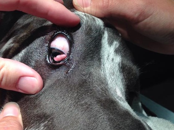
Third Eyelid Everted T Cartilage Removal
occasional the “T” shaped cartilage of the third eye lid starts to bend forward. This causes a deformation of the third eyelid resulting in irritation, and discharge to the eye. It can be easily mistaken for a prolapse of the third eye lid ( Cherry eye). When this occurs it is best the deformed cartilage be removed. It is not uncommon to have the issue develop in both eyes, and usually not at the same time.
$750 – $950
Third Eyelid Gland Removal
Removal of a prolapsed third eyelid gland is highly controversial. Some believe it increases the risk of dry eye as pets get older. In our experience we have seen few, if any, pets develop dry eye secondary to removal of the the gland alone. KCS (keratoconjunctivitis sicca) is the most common cause of dry eye in pets. This is an autoimmune disease that attacks tear production cells of the eye. So, from our experience have not seen it have a clinical significant impact on dry eye cases.
We do though encourage owners that can afford eyelid Tacking to do so. We do believe it best to allow a pet to keep its third eyelid gland when ever possible. We do recognize that some owners can not except the cost or risk of tacking failure and may elect to simply have the glands removed. We will perform the procedure only with full informed consent of the owner of the reported risk of full removal of the third eyelid glands.
$550.00
Third Eyelid Gland Tacking
Prolapse of the gland of the Third eyelid is commonly known as “Cherry eye”. The preferred method of repair is by attempting to tack the gland back into a pocket that is created on the inside of the third eyelid. Our doctors have good success at this procedure but re-prolapse is always possible.
$750 – 950.00